Research
Big picture: Human pluripotent stem cells as a model for the embryo
How an amorphous lump of identical cells is able to develop into a human being is a profound question of equal intellectual and practical importance. Its fascination springs in part from the desire to know where we come from, and how we differ from other animals. Yet the ability to direct differentiation of cultured cells into distinct cell types for therapeutic use — perhaps even coax them to form complete organs — relies on answering the same question. Ethical considerations make experimentation on human embryos unacceptable and on other mammals undesirable. Moreover, significant interspecies differences complicate inference about human development from animal models. This challenges us to find answers in alternative ways.
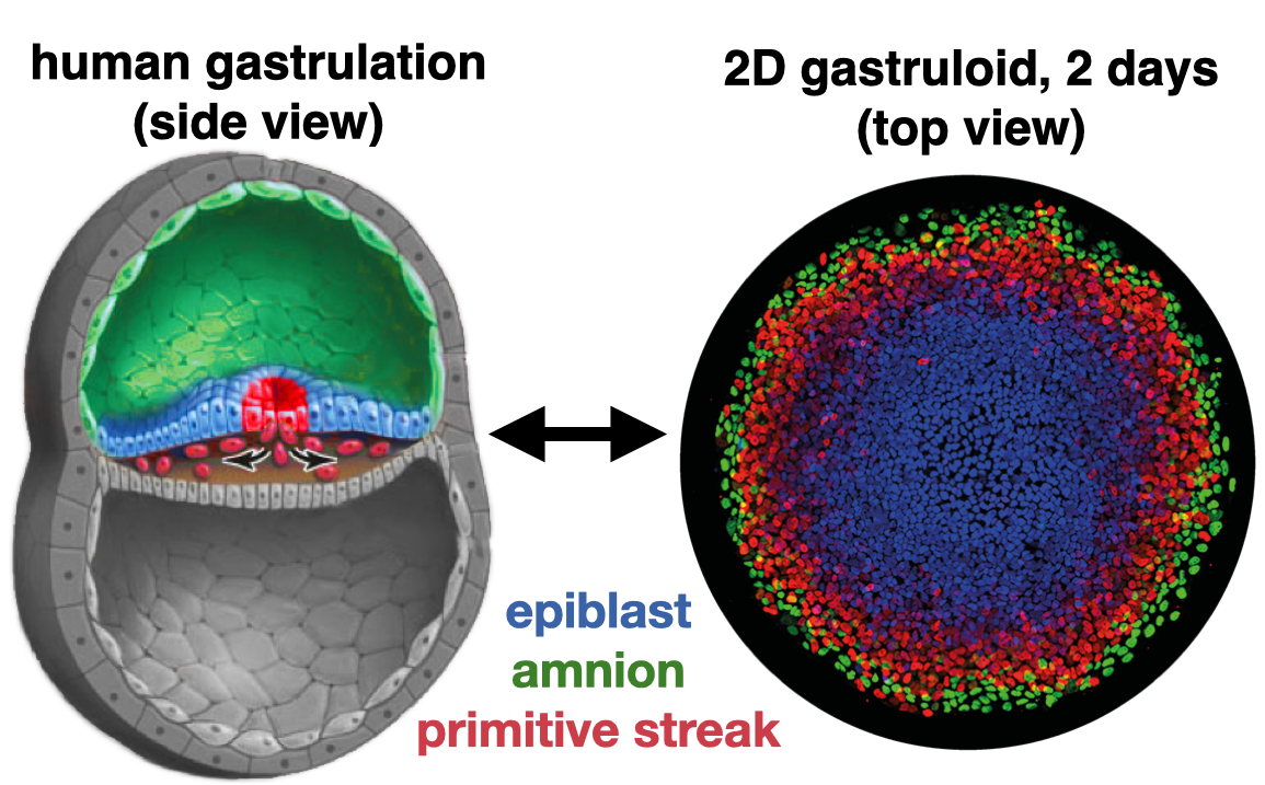
Remarkably, human pluripotent stem cells (hPSC) retain the embryo’s innate capacity to self-organize. The embryonic body plan is laid out during gastrulation, when pluripotent cells commit to one of three lineages — ectoderm, mesoderm, or endoderm — that organize into three germ layers. Upon treatment with the differentiation-inducing molecule BMP4, hPSCs confined to a disc by a micropatterned substrate mimic this process and reproducibly differentiate into concentric rings corresponding to each germ layer. These micropatterned colonies are now commonly referred to as 2D human gastruloids. Importantly, the same genetic circuitry as in the embryo orchestrates the pattern in 2D gastruloids. By systematically varying the environment we can probe underlying mechanisms with throughput and precision unachievable in living embryos.
Read more about stem cells as a model for embryonic patterning, or as a platform to study early human development, in one of our recent reviews.
Research themes
Morphogen signaling dynamics
Morphogens are diffusible signaling molecules thought to determine cell fates in a level-dependent way during developmental patterning. However, in the vertebrate embryo, cells experience changing morphogen levels during differentiation, making this static model insufficient. To address this, we developed single-cell tracking of signaling linked to cell fate, and used it to determine how cells interpret BMP signaling. We discovered that time-integrated (total) BMP signaling controls differentiation: both the level and duration of signaling activity impact cell fate only by changing the time integral of signaling, so level and duration are interchangeable in this context. In a stem cell model for embryonic patterning, we showed that the integral model correctly predicts changes in cell fate domains when signaling is perturbed. Using a screen, we found three families of transcription factors, including SOX, that may integrate BMP signaling; direct manipulation of SOX2, live-cell imaging, genomic data, and mathematical modeling support a key role for SOX2. Read our paper on time-integrated BMP signaling.
Human germ cell differentiation
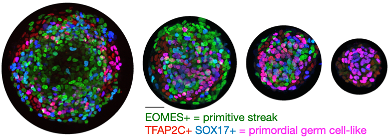
Primordial germ cell (PGC) specification is the first step in development of the germ line, and much about human PGC specification remains poorly understood because of the inaccessibility of the early post-implantation human embryo. We discovered that in 2D human gastruloids, PGC-like cells are reproducibly specified, and that on small micropatterns the fraction reaches ~70% compared with an average 30% by previous methods. This platform let us show that Wnt — previously considered the key signal in the mesoderm-versus-PGC decision — acts indirectly to induce Nodal, and that Nodal is the limiting factor. We developed a mathematical model for how the relative timing and magnitude of BMP and Nodal control the decision between amnion, primordial germ cells, and primitive streak, which may resolve the debate about whether PGC progenitors are amnion-like or mesoderm-like: it can be both. Read our paper on micropatterned differentiation of PGC-like cells and our perspective on PGC differentiation.
Human mesoderm development
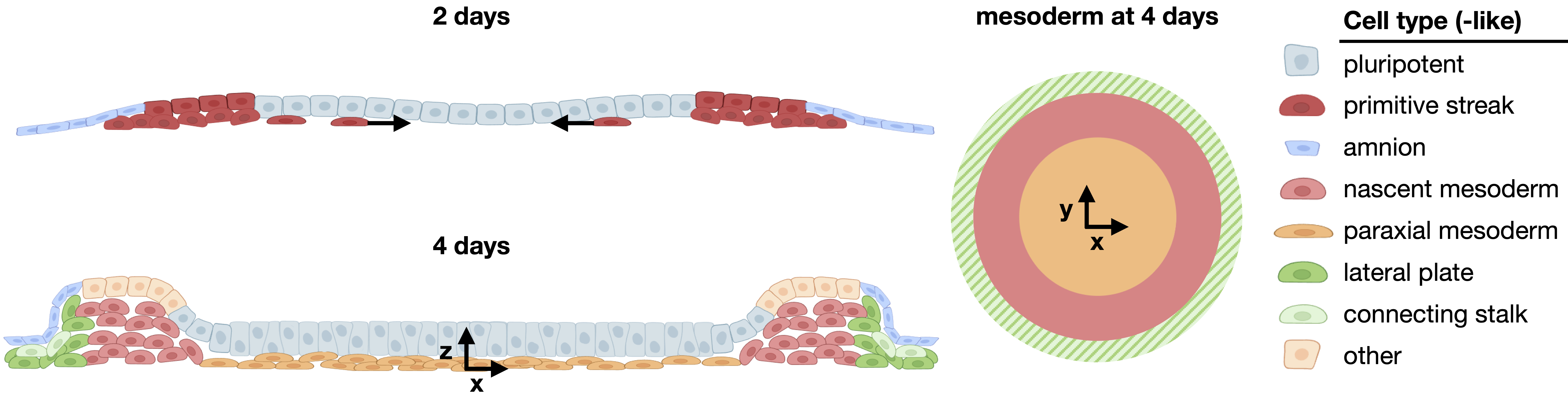
Because of their simplicity and reproducibility, 2D gastruloids are ideal for high-throughput quantitative studies of tissue patterning. However, until recently they were only studied for two days. We extended this system to four days and discovered a phase of highly reproducible morphogenesis during which directed migration from the primitive streak-like region gives rise to a mesodermal layer beneath an epiblast-like layer. Multiple types of mesoderm arise with striking spatial organization, including lateral mesoderm-like cells on the colony border and paraxial mesoderm-like cells further inside. Single-cell transcriptomics showed strong similarity to mesoderm in human and non-human primate embryos. Having established the first model of human mesoderm migration, we can now dissect the mechanisms underlying this essential and poorly understood developmental process. Check out our preprint on mesoderm patterning in human gastruloids.
Role of tissue geometry and topology in patterning
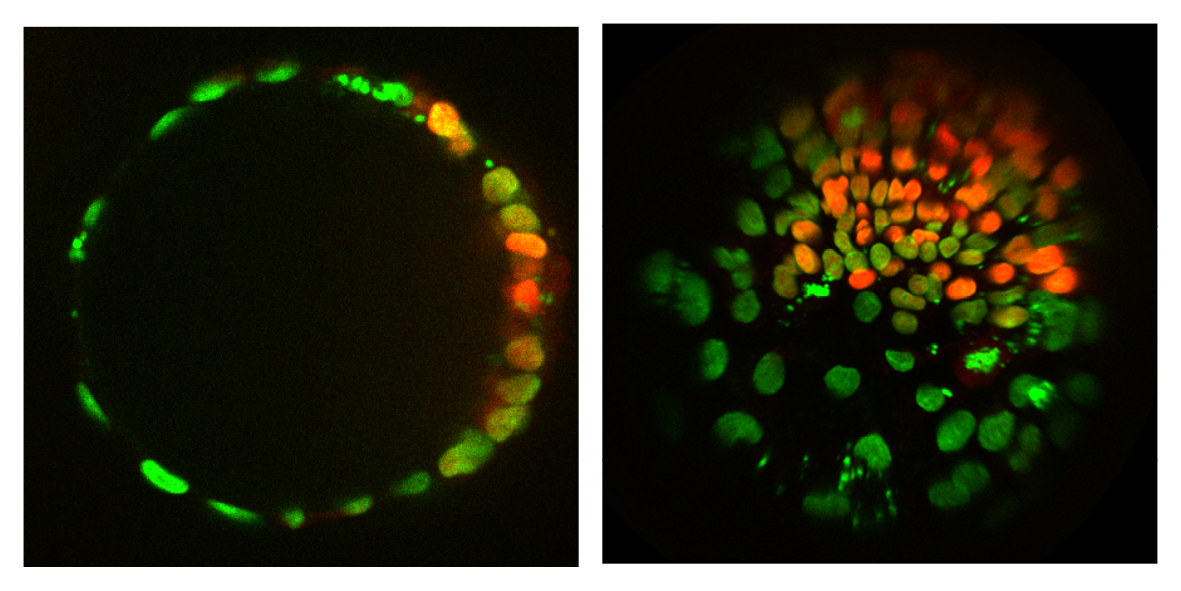
Understanding the interplay between pattern and shape, through which the body plan is established, is a major challenge in developmental biology. Unlike in embryos, the geometry (shape) and topology (how things are connected) of stem cell colonies can be controlled to study the effect on patterning. The simplest models for spatial differentiation depend on the existence of a colony border. However, by growing cells on spheres we found that patterning still occurs, suggesting the underlying mechanism is the spontaneous formation of a morphogen gradient through an activator-inhibitor system, as suggested long ago by Alan Turing. Looking at patterning on surfaces with different symmetry and shape will further constrain the mechanism of symmetry breaking and provide insight into the establishment of the body axes.